This site requires JavaScript! Please, enable it in the browser!
Home
About Scrubdin
Search
Popular Polls
Latest
Popular Polls
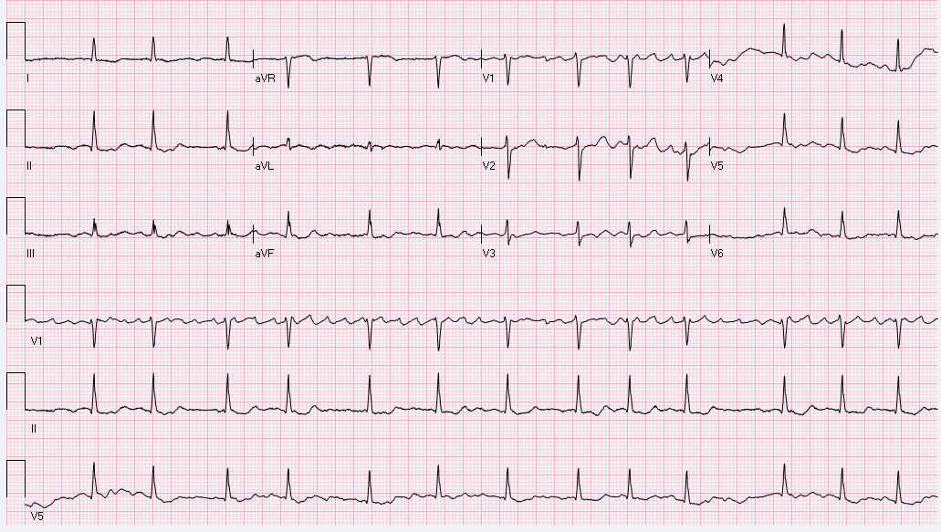
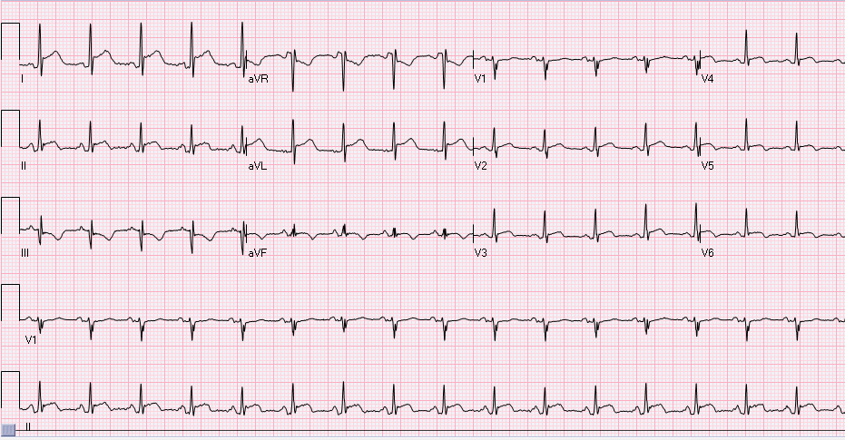
How do you describe this rhythm?
·
2025-06-17T12:37:21+0000
·
1 comments
·
Fathi Ali
1-Atrial fibrillation
2-Atrial flutter, typical
3-Atrial flutter, atypical
4-A fib / flutter
5-Sinus rhythm with artifact
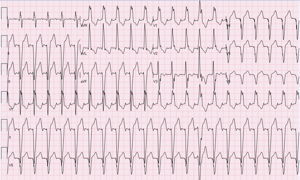
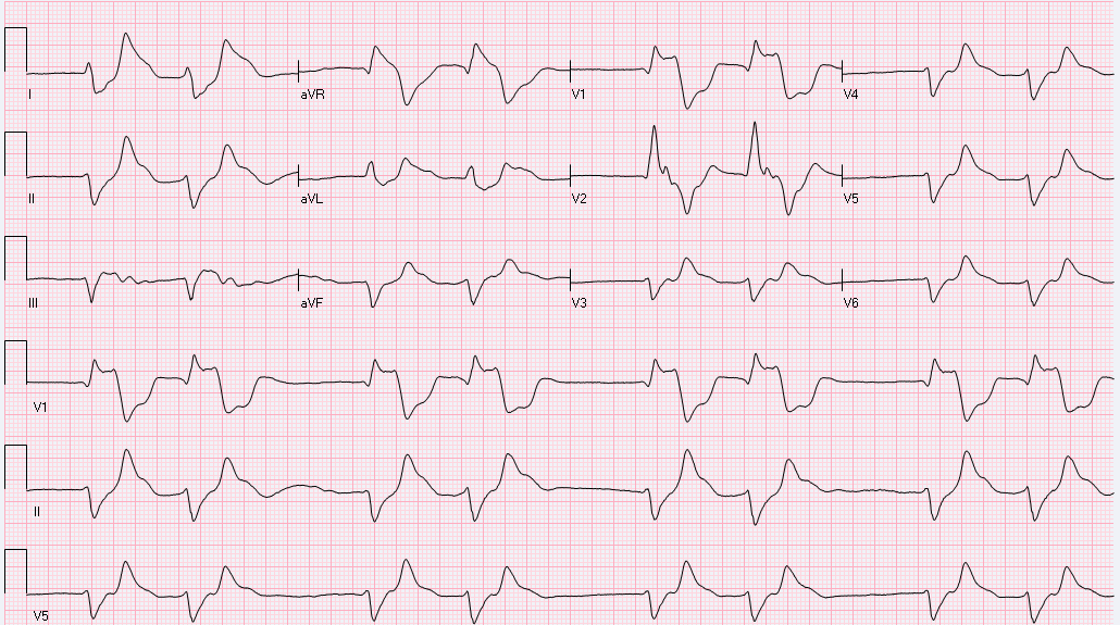
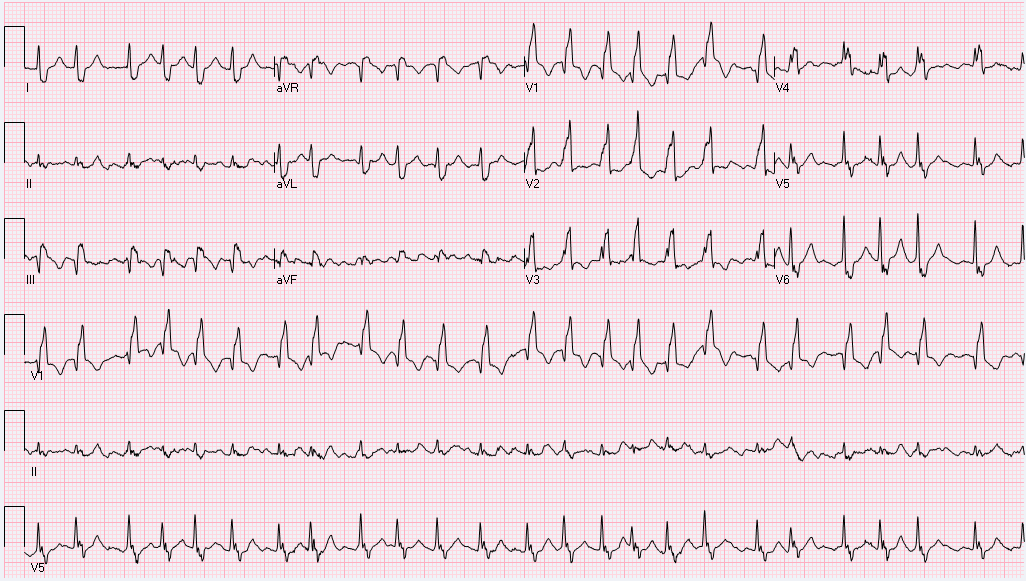
A 55-y-o male with h/o stroke. Found to be aphasic awake but non-communicating. HR: 130s, and BP: 160/80. No more history obtainable.
·
2025-06-24T18:46:52+0000
·
1 comments
·
Fathi Ali
1. A fib with RBBB – Rate control with BB/CCB.
2. Undifferentiated tachycardia – Challenge with Adenosine.
3. VT – proceed with Amiodarone or cardioversion, if needed.
4. Sinus tachycardia with RBBB – initiate on BB or calcium channel blockers.
A 46-year-old African American male presented to the ER with severe chest pain. Patient woke up in the morning complaining pressure and tightness across the chest rated 9/10. His blood...
·
2025-07-16T14:00:52+0000
·
1 comments
·
Fathi Ali
a) Proximal LAD lesion.
b) Left main disease.
c) Multivessel disease including LAD, circumflex coronary artery and right coronary artery.
d) Normal coronary arteries.
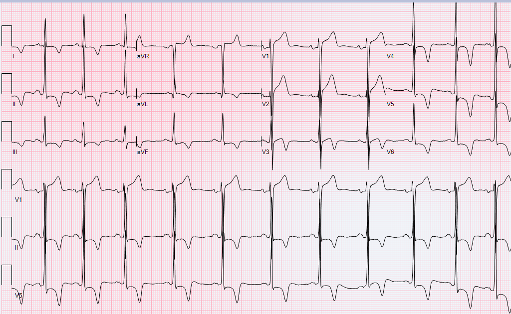
This EKG was taken from a 68-year-old male who underwent electrical cardioversion for atrial tachycardia/flutter a couple of weeks ago. He has a dual chamber PM. He is feeling well...
·
2025-07-02T20:02:06+0000
·
1 comments
·
Fathi Ali
a) PM malfunction, cardioversion likely unsuccessful. refer back to cardiology.
b) Normal PM function, cardioversion likely successful.
c) Normal PM function, but cardioversion likely unsuccessful, refer back to cardiology.
d) This EKG alone is not sufficient to tell if cardioversion was successful or not.
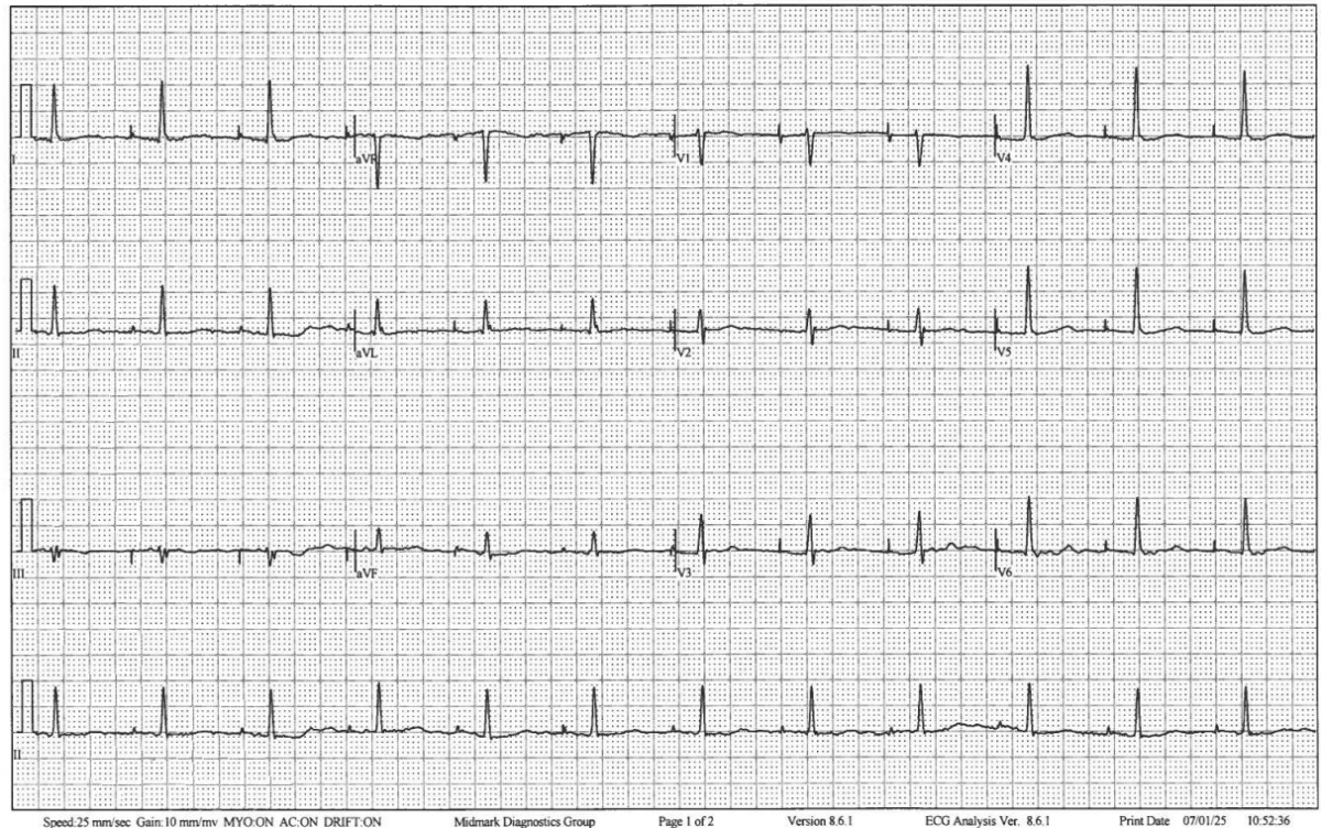
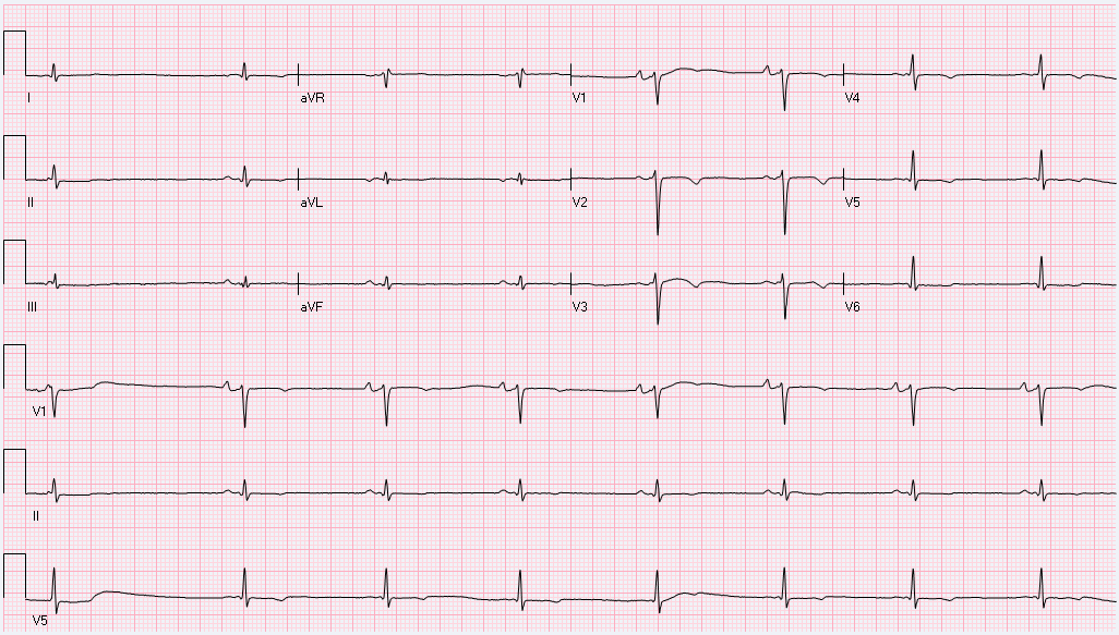
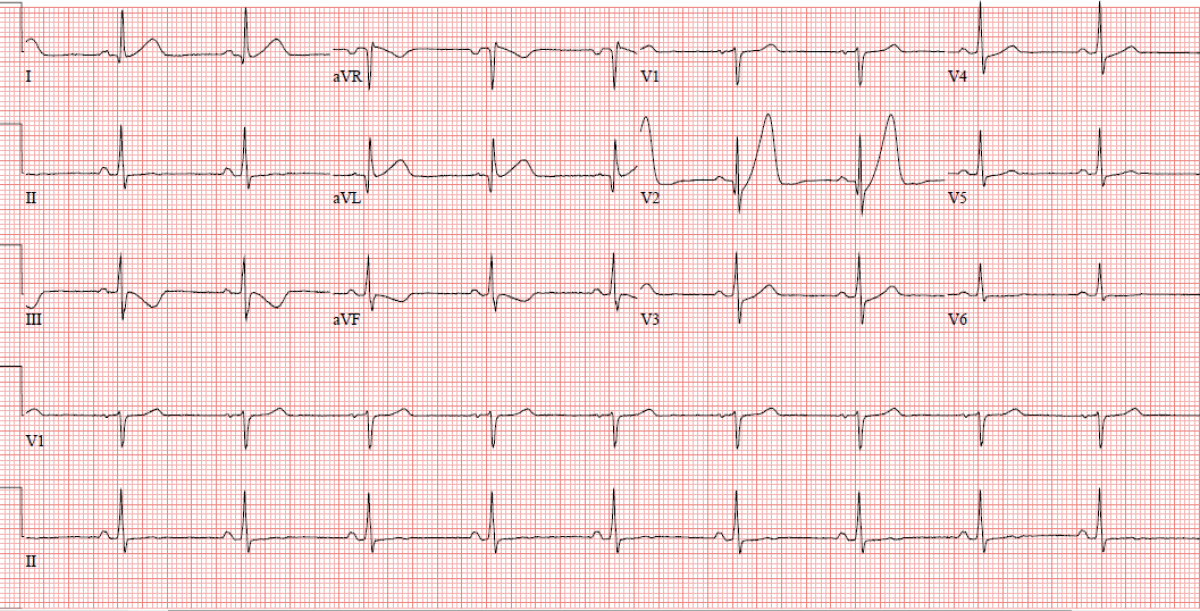
This EKG was done on an 82-year-old male who is asymptomatic. You checked his pulse in the clinic and was found to be irregular, therefore this EKG was done.Was the interpretation and next...
·
2025-05-27T16:50:31+0000
·
1 comments
·
Fathi Ali
1. Block PACs, observation.
2. Second-degree AV block, type 1, observation.
3. Second-degree AV block, type 2, observation.
4. Second-degree AV block, type 1 , pacemaker implantation.
5. Second-degree AV block, type 2, pacemaker implantation.
This EKG was taken from a 75 year-old male with paroxysmal AF. He is on longer acting metoprolol 50 mg. You noticed that his heart rate is on the slow side, but his blood pressure is normal and he...
·
2025-05-23T12:52:05+0000
·
1 comments
·
Fathi Ali
Cut back his metoprolol or stop it completely.
He is asymptomatic and some bradycardia is expected with beta-blockers, keep on metoprolol and observe.
Consider adding amiodarone at this time to minimize AF recurrence.
Consider switching metoprolol with diltiazem CD 240.
“Slow Beats, Fast Decisions: Who will Save the Day?”A 68-year-old female with past history of diabetes and hypertension as well as peripheral vascular disease with prior right limb below-knee...
·
2025-07-22T12:00:05+0000
·
1 comments
·
Fathi Ali
a. Intervention Cardiology - cardiac catheterization to address critical ischemia.
b. Nephrology -emergency hemodialysis.
c. Pulmonary and critical care - pulmonary embolus with multi organ failure.
d. Psychiatry - tricyclic antidepressant overdose.
e. Palliative care - advanced cancer.
This is a 56-y-o-Hispanic man in ER with SOB. His EKG is "classical" for what diagnosis?
·
2025-06-04T18:21:32+0000
·
1 comments
·
Fathi Ali
Severe COPD
Acute/subacute pulmonary embolus.
Cardiomyopathy due to Amyloidosis
Hypokalemia
None of the above
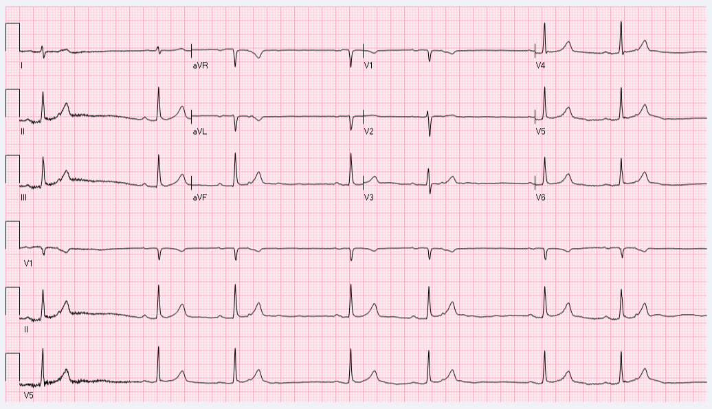
This is a 65-year-old male, otherwise healthy who presented to the ED with chest pain and was found to have elevated troponin around 622 ng/L (high sensitivity troponin).Based on the EKG findings,...
·
2025-07-09T17:48:27+0000
·
1 comments
·
Fathi Ali
a. Pericarditis, anti-inflammatory.
b. NSTEMI, Heparin, ASA, Cath later.
c. STEMI, Heparin, ASA, Cath immediately.
d. Pulmonary embolus, Heparin, ICU admission.
e. Early repolarization pattern, investigate other causes of elevaed troponin.
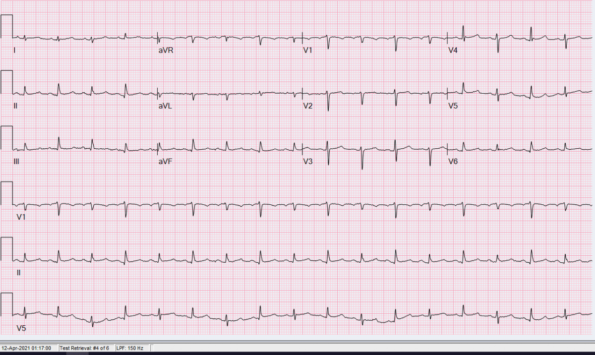
This EKG was taken from an 83-year-old female in the hospital with UTI. She reports shortness breath and palpitations. What is the rhythm interpretation?
·
2025-06-09T13:01:23+0000
·
1 comments
·
Fathi Ali
Atrial flutter with right bundle-branch block.
Ventricular tachycardia of right bundle-branch block morphology.
A Fib with right bundle-branch block.
A Fib with left bundle-branch block.
Ventricular tachycardia of left bundle-branch block morphology.
Focal STEMI!A 61 year-old-male with family h/o CAD. Presented to ED with 30 min of left sided chest pain radiates to the left shoulder. EKG on arrival shown.Initial hs-cTnT: 7 and 1-hr later...
·
2025-09-09T12:42:13+0000
·
1 comments
·
Fathi Ali
Proximal left anterior descending artery (LAD)
First Diagonal branch
Large obtuse marginal branch
Right coronary artery
This EKG was done on a 77 y-o female who was referred because of irregular heart beats. She is asymptomatic. Her PMH included HTN, and DM.Based on this EKG, what is the next step in managing this...
·
2025-05-21T12:39:35+0000
·
1 comments
·
Fathi Ali
Initiate low-dose BB and anticoagulation.
Initiate anticoagulation only.
Repeat the EKG.
Consider cardioversion after 4 weeks of anticoagulation.
Start baby aspirin and observation.
1-12
Search
Close
Home
About Scrubdin
Search
OK
Are you sure?
Yes
No
Please, enter a value here
OK
Cancel