

84 Year old lady presented to the hospital with recurrent stroke. Patient was in other hospital 8 days before because of stroke. The brain CT showed embolic stroke picture. The Echo report from the prior stroke was normal. This is her echo this admission. What are the differential diagnosis for this?
Comments
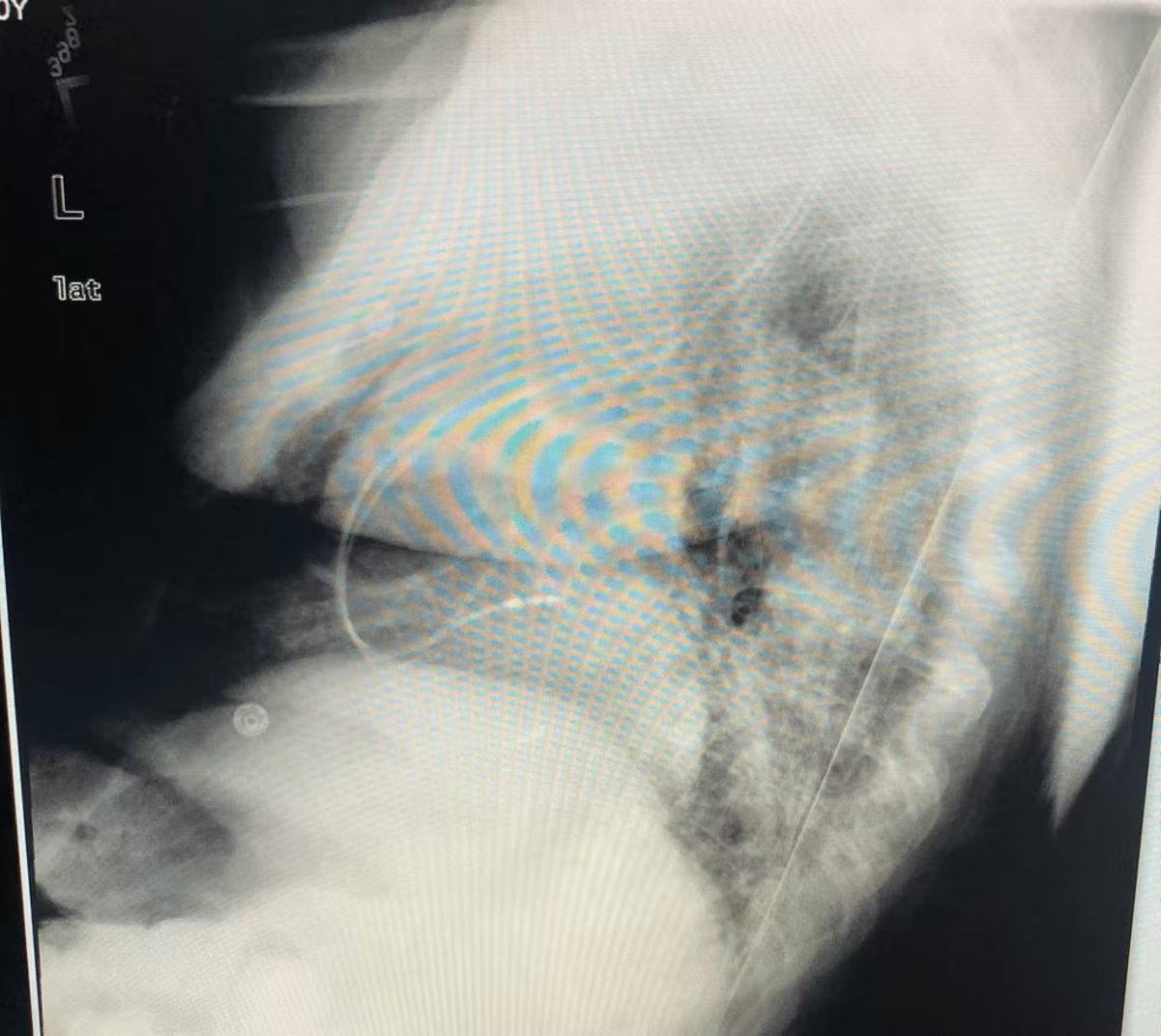
We did Cardiac CT for further assessment and the images is here
All good D/D.
The best way is to have clear TEE images, carefully assessing the mass and its origin.
Cardiac MRI might help as well.
Also there is significant MR which is suggestive of the MV involvement. Not enough images.
I have seen similar case and TEE showed rupture papillary muscle which was miss-diagnosed by TTE. That was confirmed during cardiac surgery and histopathology.
F
Nice. Thanks Never seen one, or probably seen it but didn't recognize it.
I googled it a bit, and I should say although the case sounds suspicious, but the image quality are not classical with central liquefaction and that mass on one of the views seems to be a bit far from the annulus itself. Would be nice if there is a TEE that pinpoint that.
How did you treat this patient? Also, I didn't understand the relationship of the header image (with CXR of PM) with the case!
Great case @Mohsin Salih and thanks for the challenging and very interesting and educational case.
Also, I will share a couple reviewed that I found on this. Thanks again @Mohsin Salih and the rest of the group for the nice and educational discussion.
https://scrubdin.net/view-file/cmac-review-2021-pdf
https://scrubdin.net/view-file/cmac-jacc-editorial-2021-pdf
Excellent education case. Thank you Moh! for sharing the case. Did you keep the patient on anticoagulant?
F
Nice illustration, thanks @Mansour Khaddr !
great case but not fully convinced
I don't see the rupture point
can we have more of the CT in the short axis slices