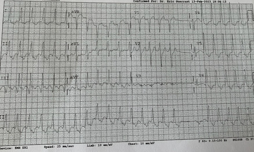
Personally I will call this wide complex tachycardia (using lead III QRS duration around 120 ms).
heart rate around 150 bpm
axis is normal
Atrial activity seen after each QRS complex except the initial beat after the conducted sinus beat at the end of the rhythm strip.
the atrial activity has positive morphology in lead aVR and negative in inferior leads which means the direction of the electrical activity from inferior to superior (ventricular tachycardia with VA conduction/atrial tachycardia/junctional or AVNRT ….)
Unlikely atrial tachycardia given the flat line at the termination and also no atrial activity seen prior to initiation.
certainly not atrial flutter.
I don’t see delta wave in the conducted sinus beat.
I will go with ventricular tachycardia (probably septal vt)
also, the morphology of the initial r wave in V2 is more than 40 ms (not typical for LBBB) goes with VT.
overall:
Ventricular tachycardia with VA 1:1 conduction except for one beat after the conducted sinus beat (? AV node Refractory after conducted sinus beat)


VT
F
Why?
Personally I will call this wide complex tachycardia (using lead III QRS duration around 120 ms).
heart rate around 150 bpm
axis is normal
Atrial activity seen after each QRS complex except the initial beat after the conducted sinus beat at the end of the rhythm strip.
the atrial activity has positive morphology in lead aVR and negative in inferior leads which means the direction of the electrical activity from inferior to superior (ventricular tachycardia with VA conduction/atrial tachycardia/junctional or AVNRT ….)
Unlikely atrial tachycardia given the flat line at the termination and also no atrial activity seen prior to initiation.
certainly not atrial flutter.
I don’t see delta wave in the conducted sinus beat.
I will go with ventricular tachycardia (probably septal vt)
also, the morphology of the initial r wave in V2 is more than 40 ms (not typical for LBBB) goes with VT.
overall:
This is a parahisian VT. VT arising around the His area in the upper ventricular septal area.
Thank you for sharing this case with us Dr Yousef!!
F
@Yousef Darrat thanks for this nice case. Can you kindly educate us about Parasisian VT by listing some brief note here?