Dear colleagues/friends,
I need your help with a case that we saw last week. I am not sure we know exactly what is going on with her. I am wondering if you see a similar case before, or how would you manage her if she is yours. Will tell you later what we decided to do.
Appreciate your help!
tnx
History:
66-year-old female with PMH significant for hypertension, type 2 diabetes mellitus, and chronic pain (followed at pain clinic) presents to ED for evaluation of chest pain.
Patient reports intermittent sharp, achy chest pain beginning 3 days ago. Mostly located above and around left breast with some radiation into left arm. She also reports mid upper back pain at times. Exacerbated with deep breaths but denies alleviating factors.
She denies prior hx of DVT/PE, recent prolonged travel, recent surgeries. Denies fever, chills, palpitations.
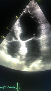
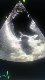
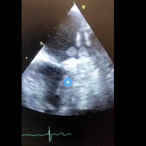
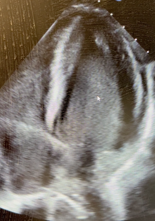
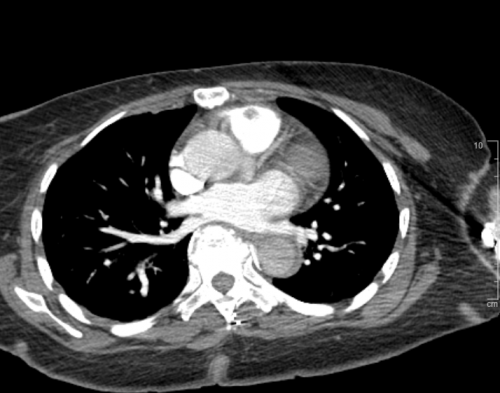
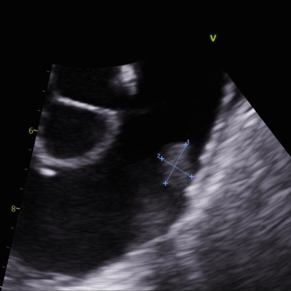
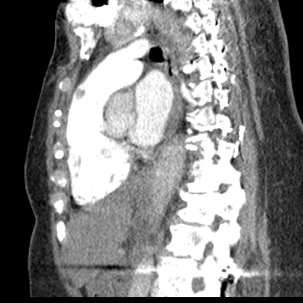
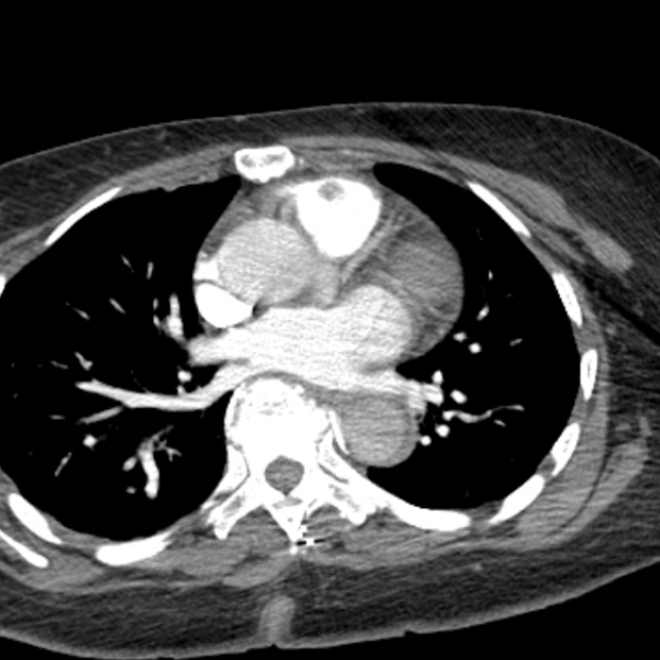
ED workup mostly unremarkable but D-Dimer is elevated at 1580. Chest x-ray negative for acute process. CT a chest showed filling defect pulmonic valve. Questionable for focal thrombus, potentially infected vegetation, or soft tissue mass/neoplasm.
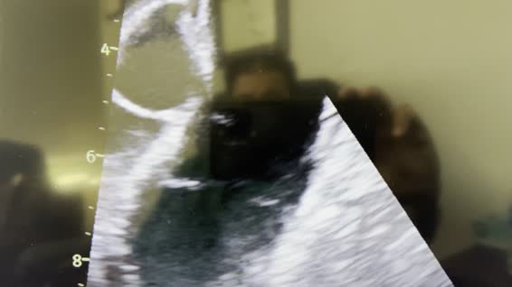
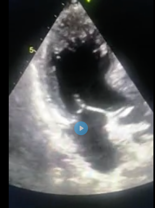
Attached images from CT (with contrast) and TEE.

An 80-year-old male presented with persistent right shoulder pain, achy in nature for the last couple of weeks. No significant improvement post local cortisone injection.
PMH: CAD s/p 4V CABG (grafts occluded except RCA graft). Coronary/graft angiogram in 5/2022 performed for similar symptoms showed RCA vein graft ostial 95% stenosis s/p PCI with resolution of his shoulder pain. Also, had balloon angioplasty only to proximal LAD in-stent restenosis as already has 2 stents at that location. He was doing well till 4-6 weeks ago when his right shoulder pain recurred. He also has CKD stage IV, PVD, carotid artery disease s/p intervention and ischemic CVA with no obvious residual weakness.
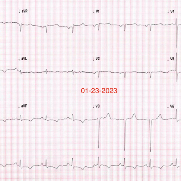
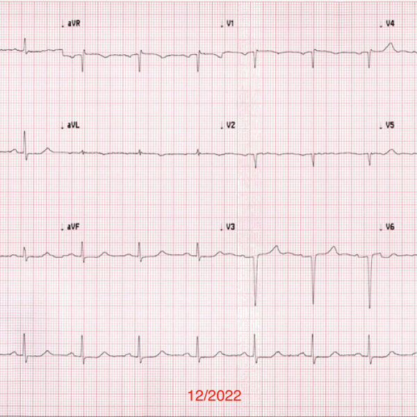
His initial EKG from 12/2022 and second EKG done this week. Also, limited echo images posted. His echo in 4/2022 showed LVEF 55% with no regional wall motion abnormality.
It is a bread and butter case. What is the echocardiogram findings?